1. Actinomycosis
① Definition
Actinomycosis is an infectious disease caused by anaerobic or microaerophilic bacteria that normally reside in the oral cavity, colon, and female genital tract.
② Pathogenesis
The disease occurs when the mucosal barrier is disrupted, allowing bacteria to invade deeper tissues.
As the infection progresses, it forms granulomatous tissue, extensive reactive fibrosis, necrosis, abscesses, and fistulae.
③ Characteristics
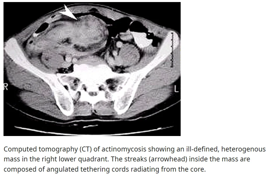
- It tends to spread without regard to anatomical barriers.Formation of sulfur granules.
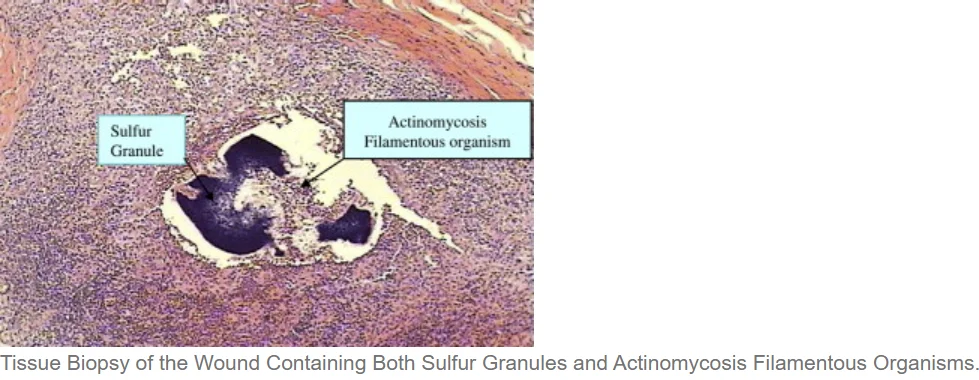
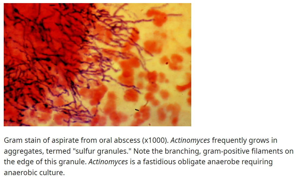
- These are yellow granules found in pus. The name is misleading because they are not composed of sulfur but rather aggregates of immune cells and bacterial colonies.
- They represent Actinomyces colonies, typically surrounded by an eosinophilic granulation tissue zone.
- However, not all Actinomyces species form sulfur granules.
- Example: In cases like tonsillar infection, where surrounding tissue reaction is minimal, sulfur granules may not be formed.
④ Risk Factors
- Diabetes mellitus (14%)
- Immunosuppression (14%)
- Malnutrition, local tissue damage, or inflammation (71%)
- Examples: Trauma, dental procedures, gingivitis, radiation therapy, osteonecrosis, poor oral hygiene.
Clinical Features and Diagnosis
① Infection Progression
- Chronic and slowly progressive infection.
② Diagnostic Challenges
- Symptoms may take weeks to months to appear after infection.
- Time to diagnosis varies depending on specimen type and incubation period required for bacterial recovery.
③ Primary Sites of Infection and Symptoms


- Orocervicofacial region (40–60%)
- Common symptoms: Pain, trismus (difficulty opening the mouth).
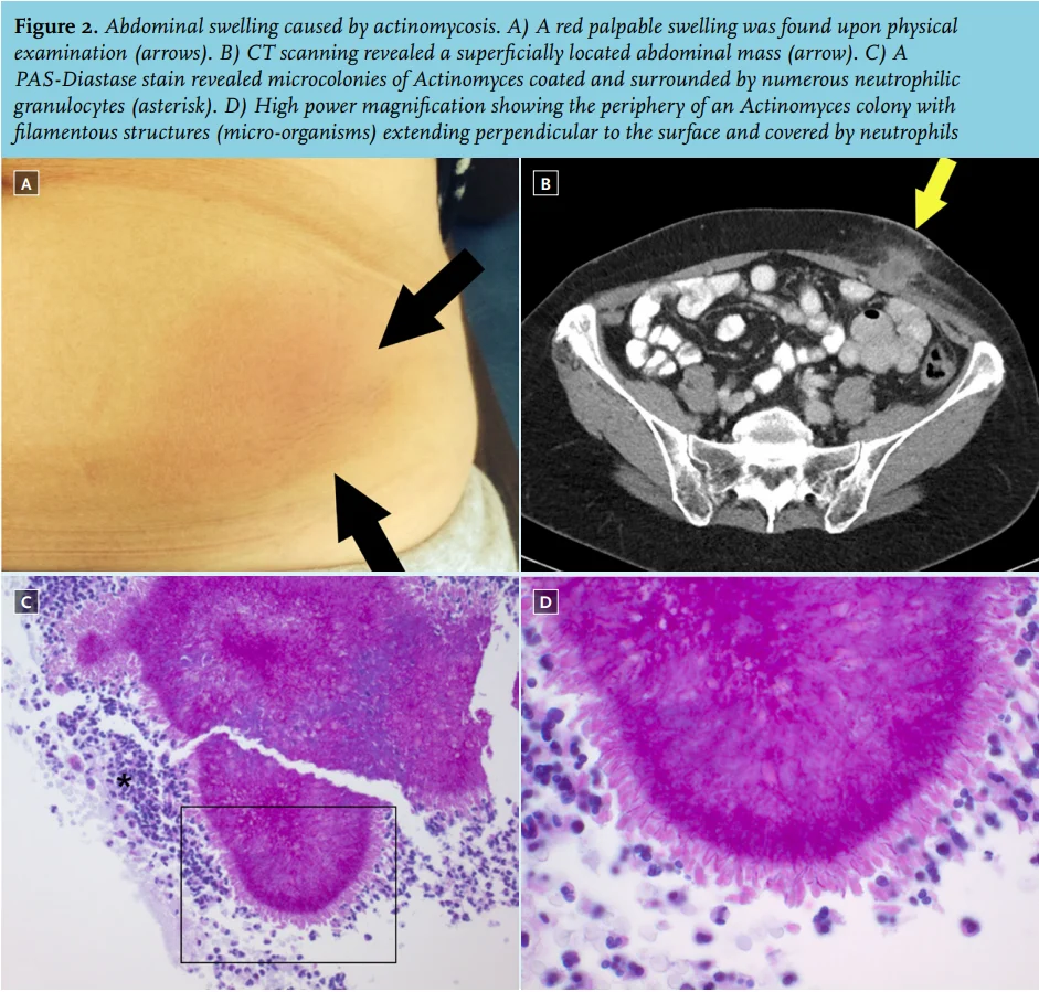
- Abdominopelvic region (25–35%)
- Occurs after abdominal surgery, trauma, neoplasm, or perforation.
- Symptoms: Fever, fatigue, weight loss, chronic abdominal pain.
- CT scan: Non-specific findings, usually showing a solid mass adjacent to affected structures.
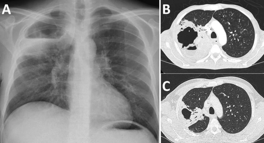
- Thoracopulmonary region (20–25%)
- Symptoms: Fever, weight loss, dyspnea, chest pain, severe cough, hemoptysis.
- CT scan: Cavity formation, mass-like lesions, inflammation, and pleural involvement.
- May mimic lung cancer.
- Cutaneous form (3–5%)
④ Diagnostic Procedures
- Obtain diagnostic specimens.
- Blood cultures and fluid or tissue samples from masses, fistulae, or sinuses should be sent to microbiology and pathology laboratories.
- Final diagnosis requires pathogen isolation and identification via culture or histopathology.
3. Treatment
① Severe or Extensive Actinomycosis
- Intravenous Penicillin G (10–20 million units daily, divided every 4–6 hours) is recommended initially.
- Ceftriaxone (1–2 g every 24 hours) is an alternative for outpatient treatment.
- Intravenous therapy generally lasts 4–6 weeks before transitioning to oral therapy.
- Surgical intervention may be necessary in some severe cases.
Reference
- UpToDate: Actinomycosis. Available at: uptodate.com