1. Fracture Healing Steps

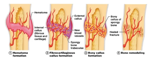
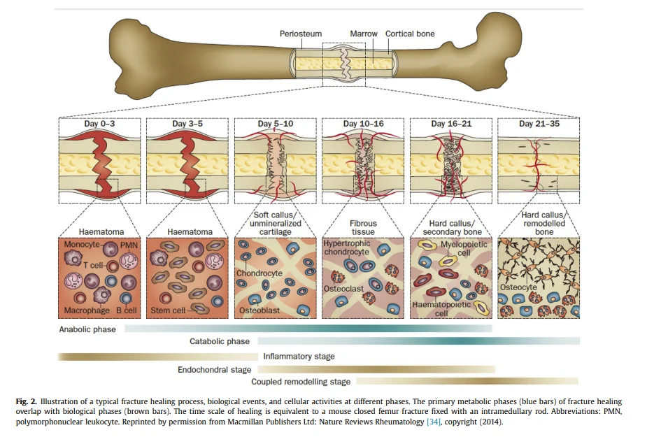
1) Hematoma Formation (Days 1-5)
- Initiates immediately after the fracture.
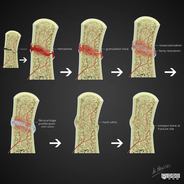
- Blood vessels supplying the bone and periosteum rupture, forming a hematoma around the fracture site.
- The hematoma coagulates, creating a temporary scaffold for subsequent healing.
- Bone injury triggers the release of pro-inflammatory cytokines such as TNF-α, bone morphogenetic proteins (BMPs), and interleukins (IL-1, IL-6, IL-11, IL-23).
- These cytokines recruit macrophages, monocytes, and lymphocytes to stimulate essential cellular activities in the fracture site.
- These immune cells work together to remove necrotic tissue and secrete vascular endothelial growth factor (VEGF) to promote healing.
2) Fibrocartilaginous Callus Formation (Days 5-11)
- VEGF release stimulates angiogenesis in the fracture site, leading to the development of fibrin-rich granulation tissue within the hematoma.
- Additional mesenchymal stem cells (MSCs) are recruited and begin differentiating into fibroblasts, chondroblasts, and osteoblasts.
- Cartilage formation begins, leading to the development of a collagen-rich fibrocartilaginous network.
3) Bony Callus Formation (Days 11-28)
- The cartilage callus undergoes endochondral ossification.
- RANK-L expression promotes the differentiation of chondroblasts, chondrocytes, osteoblasts, and osteoclasts.
- The cartilaginous callus is progressively resorbed and begins to mineralize.
- Newly formed blood vessels continue to proliferate, allowing further migration of MSCs.
- By the end of this stage, a hard, calcified immature bone structure is established.
4) Bone Remodeling (Day 18 onward, lasting months to years)
- Ongoing osteoblast and osteoclast activity results in continuous remodeling of the hard callus through “coupled remodeling.”
- Coupled remodeling refers to a balanced process of resorption by osteoclasts and new bone formation by osteoblasts.
- The central portion of the callus is gradually replaced by cortical bone, while the peripheral edges transform into lamellar bone.
- Bone remodeling continues for months, ultimately restoring the normal bone structure.


2. Definition of Fracture Union
1) Average Healing Time for Common Fractures
Upper Extremity:
- Phalanges: ~3 weeks
- Metacarpals: 4-6 weeks
- Distal radius: 4-6 weeks
- Humerus: 6-8 weeks
- Forearm: 8-10 weeks
Lower Extremity:
- Metatarsals: >6 weeks
- Tibia: ~10 weeks
- Femoral neck and femoral shaft: ~12 weeks
2) Most Common Clinical Criteria for Fracture Union
- Absence of pain or tenderness on weight-bearing (49%)
- No pain or tenderness on palpation or physical examination (39%)
- Ability to bear weight (18%)
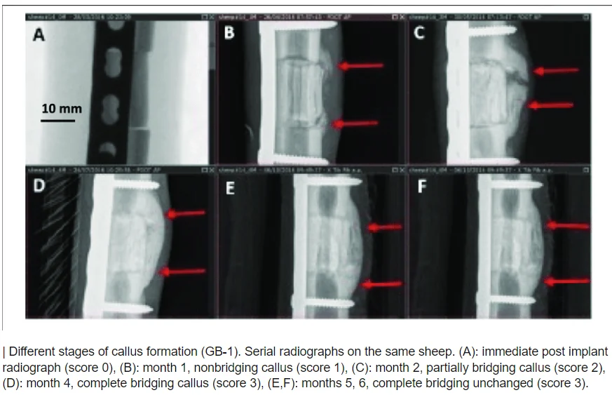
3) Radiological Criteria for Fracture Union
- Fracture site connection through bone, callus, and trabeculae
- Presence of three bridging calluses on AP or lateral X-ray views
- Disappearance of the fracture line
Note: Callus formation is a key indicator of bone healing.


References
- UpToDate.com
- J Orthop Trauma 2010;24:S76–S80
- NIH – Fracture Healing Overview – Jonathon R. Sheen; Vishnu V. Garla.
- https://radiopaedia.org/articles/fracture-healing